Figure 2
Pulmonary mucormycosis in post-pulmonary tuberculosis as an emerging risk factor: A rare case report
Divya Khanduja* and Naveen Pandhi
Published: 30 July, 2021 | Volume 5 - Issue 1 | Pages: 059-063
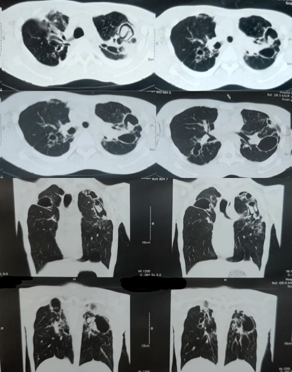
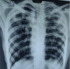
Figure 2:
CECT Chest showed multifocal areas of cavitation involving bilateral upper and lower lobes of lungs. Fungal balls in bilateral upper lobe cavities showing air crescent sign.
Read Full Article HTML DOI: 10.29328/journal.jprr.1001026 Cite this Article Read Full Article PDF
More Images
Similar Articles
-
Epidemiological Prevalence of Tuberculosis in the State of Maranhão between 2014 and 2016Pedro Agnel Dias Miranda Neto*,Hortência Biatriz de Melo Santana, Vanessa Maria das Neves,Hermerson Sousa Maia,Thayná Milena Nunes França,Rosana Karla Costa. Epidemiological Prevalence of Tuberculosis in the State of Maranhão between 2014 and 2016. . 2019 doi: 10.29328/journal.jprr.1001012; 3: 013-016
-
Pulmonary mucormycosis in post-pulmonary tuberculosis as an emerging risk factor: A rare case reportDivya Khanduja*,Naveen Pandhi. Pulmonary mucormycosis in post-pulmonary tuberculosis as an emerging risk factor: A rare case report. . 2021 doi: 10.29328/journal.jprr.1001026; 5: 059-063
-
Long-term results of 10 years of observation of cured cases of pulmonary tuberculosisBobokhojaev OI*. Long-term results of 10 years of observation of cured cases of pulmonary tuberculosis. . 2022 doi: 10.29328/journal.jprr.1001036; 6: 007-011
-
Efficiency of Artificial Intelligence for Interpretation of Chest Radiograms in the Republic of TajikistanBobokhojaev OI*,Abdulloev NN,Khushvakhtov ShD,Shukurov SG. Efficiency of Artificial Intelligence for Interpretation of Chest Radiograms in the Republic of Tajikistan. . 2024 doi: 10.29328/journal.jprr.1001064; 8: 069-073
Recently Viewed
-
Obesity in Patients with Chronic Obstructive Pulmonary Disease as a Separate Clinical PhenotypeDaria A Prokonich*, Tatiana V Saprina, Ekaterina B Bukreeva. Obesity in Patients with Chronic Obstructive Pulmonary Disease as a Separate Clinical Phenotype. J Pulmonol Respir Res. 2024: doi: 10.29328/journal.jprr.1001060; 8: 053-055
-
Current Practices for Severe Alpha-1 Antitrypsin Deficiency Associated COPD and EmphysemaMJ Nicholson*, M Seigo. Current Practices for Severe Alpha-1 Antitrypsin Deficiency Associated COPD and Emphysema. J Pulmonol Respir Res. 2024: doi: 10.29328/journal.jprr.1001058; 8: 044-047
-
Navigating Neurodegenerative Disorders: A Comprehensive Review of Current and Emerging Therapies for Neurodegenerative DisordersShashikant Kharat*, Sanjana Mali*, Gayatri Korade, Rakhi Gaykar. Navigating Neurodegenerative Disorders: A Comprehensive Review of Current and Emerging Therapies for Neurodegenerative Disorders. J Neurosci Neurol Disord. 2024: doi: 10.29328/journal.jnnd.1001095; 8: 033-046
-
Metastatic Brain Melanoma: A Rare Case with Review of LiteratureNeha Singh,Gaurav Raj,Akshay Kumar,Deepak Kumar Singh,Shivansh Dixit,Kaustubh Gupta*. Metastatic Brain Melanoma: A Rare Case with Review of Literature. J Radiol Oncol. 2025: doi: 10.29328/journal.jro.1001080; 9: 050-053
-
Validation of Prognostic Scores for Attempted Vaginal Delivery in Scar UterusMouiman Soukaina*,Mourran Oumaima,Etber Amina,Zeraidi Najia,Slaoui Aziz,Baydada Aziz. Validation of Prognostic Scores for Attempted Vaginal Delivery in Scar Uterus. Clin J Obstet Gynecol. 2025: doi: 10.29328/journal.cjog.1001185; 8: 023-029
Most Viewed
-
Evaluation of Biostimulants Based on Recovered Protein Hydrolysates from Animal By-products as Plant Growth EnhancersH Pérez-Aguilar*, M Lacruz-Asaro, F Arán-Ais. Evaluation of Biostimulants Based on Recovered Protein Hydrolysates from Animal By-products as Plant Growth Enhancers. J Plant Sci Phytopathol. 2023 doi: 10.29328/journal.jpsp.1001104; 7: 042-047
-
Sinonasal Myxoma Extending into the Orbit in a 4-Year Old: A Case PresentationJulian A Purrinos*, Ramzi Younis. Sinonasal Myxoma Extending into the Orbit in a 4-Year Old: A Case Presentation. Arch Case Rep. 2024 doi: 10.29328/journal.acr.1001099; 8: 075-077
-
Feasibility study of magnetic sensing for detecting single-neuron action potentialsDenis Tonini,Kai Wu,Renata Saha,Jian-Ping Wang*. Feasibility study of magnetic sensing for detecting single-neuron action potentials. Ann Biomed Sci Eng. 2022 doi: 10.29328/journal.abse.1001018; 6: 019-029
-
Pediatric Dysgerminoma: Unveiling a Rare Ovarian TumorFaten Limaiem*, Khalil Saffar, Ahmed Halouani. Pediatric Dysgerminoma: Unveiling a Rare Ovarian Tumor. Arch Case Rep. 2024 doi: 10.29328/journal.acr.1001087; 8: 010-013
-
Physical activity can change the physiological and psychological circumstances during COVID-19 pandemic: A narrative reviewKhashayar Maroufi*. Physical activity can change the physiological and psychological circumstances during COVID-19 pandemic: A narrative review. J Sports Med Ther. 2021 doi: 10.29328/journal.jsmt.1001051; 6: 001-007

HSPI: We're glad you're here. Please click "create a new Query" if you are a new visitor to our website and need further information from us.
If you are already a member of our network and need to keep track of any developments regarding a question you have already submitted, click "take me to my Query."


