Figure 2
Spontaneous pneumomediastinum: case report
Pedrós García Sergio, Yakasova Elena, Roquet-Jalmar Saus María, Gutiérrez González Nuria and Mercedes Noboa Edwin*
Published: 28 January, 2022 | Volume 6 - Issue 1 | Pages: 001-003

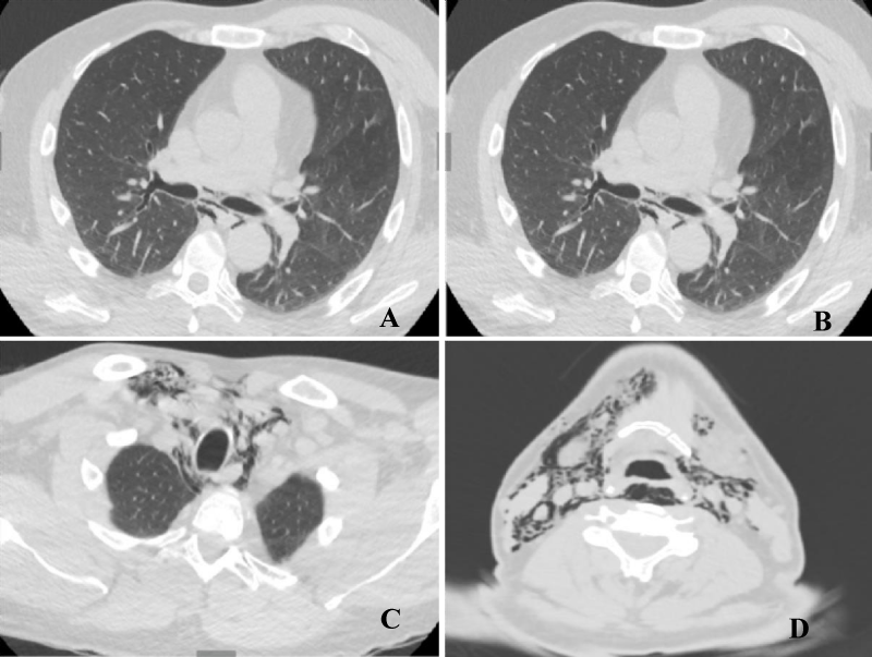
Figure 2:
Computed tomography. A: axial slice in lung window, shows air in the middle mediastinum surrounding main bronchi, esophagus, and hemiacigus vein. B: coronal slice in lung window, where pneumomediastinum is observed without pneumothorax. Emphysema of the soft tissues of the upper mediastinum and neck with aerial dissection of the muscular and vascular structures (C and D).
Read Full Article HTML DOI: 10.29328/journal.jprr.1001034 Cite this Article Read Full Article PDF
More Images
Similar Articles
-
Fluticasone furoate/Vilanterol 92/22 μg once-a-day vs Beclomethasone dipropionate/Formoterol 100/6 μg b.i.d. in asthma patients: a 12-week pilot studyClaudio Terzano*,Francesca Oriolo. Fluticasone furoate/Vilanterol 92/22 μg once-a-day vs Beclomethasone dipropionate/Formoterol 100/6 μg b.i.d. in asthma patients: a 12-week pilot study . . 2017 doi: 10.29328/journal.jprr.1001004; 1: 013-022
-
Eosinophilic otitis media and eosinophilic asthma: Shared pathophysiology and response to anti-IL5Marieke T Drijver-Messelink*,Mariette Wagenaar,Jacqueline van der Meij,Anneke ten Brinke. Eosinophilic otitis media and eosinophilic asthma: Shared pathophysiology and response to anti-IL5 . . 2019 doi: 10.29328/journal.jprr.1001011; 3: 009-012
-
Dysfunctional breathing in childrenAmanda MP Trompenaars#,Aalt PJ Van Roest#,Anja APH Vaessen-Verberne*. Dysfunctional breathing in children. . 2020 doi: 10.29328/journal.jprr.1001013; 4: 001-005
-
Spontaneous pneumomediastinum associated with COVID-19: Rare complication of 2020 pandemicArnaldo A Nieves-Ortiz*,Vanessa Fonseca-Ferrer, Kyomara Hernández-Moya,Keren Mendez Ramirez,Jose Ayala-Rivera,Marangely Delgado, Juan Garcia-Puebla,Rosangela Fernández-Medero,Ricardo Fernandez-Gonzalez. Spontaneous pneumomediastinum associated with COVID-19: Rare complication of 2020 pandemic. . 2020 doi: 10.29328/journal.jprr.1001016; 4: 018-020
-
A Case series on Asthma-COPD overlap (ACO) is independent from other chronic obstructive diseases (COPD and Asthma)Divya Khanduja*, Naveen Pandhi. A Case series on Asthma-COPD overlap (ACO) is independent from other chronic obstructive diseases (COPD and Asthma). . 2021 doi: 10.29328/journal.jprr.1001025; 5: 054-058
-
Asthma and pregnancy prevalence in a developing country and their mortality outcomesRaul Aguilar*,Jorge Martinez,Edgar Turcios,Victor Castro. Asthma and pregnancy prevalence in a developing country and their mortality outcomes. . 2021 doi: 10.29328/journal.jprr.1001031; 5: 088-093
-
Spontaneous pneumomediastinum: case reportPedrós García Sergio,Yakasova Elena,Roquet-Jalmar Saus María,Gutiérrez González Nuria,Mercedes Noboa Edwin*. Spontaneous pneumomediastinum: case report. . 2022 doi: 10.29328/journal.jprr.1001034; 6: 001-003
Recently Viewed
-
Cystoid Macular Oedema Secondary to Bimatoprost in a Patient with Primary Open Angle GlaucomaKonstantinos Kyratzoglou*,Katie Morton. Cystoid Macular Oedema Secondary to Bimatoprost in a Patient with Primary Open Angle Glaucoma. Int J Clin Exp Ophthalmol. 2025: doi: 10.29328/journal.ijceo.1001059; 9: 001-003
-
Sex after Neurosurgery–Limitations, Recommendations, and the Impact on Patient’s Well-beingMor Levi Rivka*, Csaba L Dégi. Sex after Neurosurgery–Limitations, Recommendations, and the Impact on Patient’s Well-being. J Neurosci Neurol Disord. 2024: doi: 10.29328/journal.jnnd.1001099; 8: 064-068
-
Physiotherapy Undergraduate Students’ Perception About Clinical Education; A Qualitative StudyPravakar Timalsina*,Bimika Khadgi. Physiotherapy Undergraduate Students’ Perception About Clinical Education; A Qualitative Study. J Nov Physiother Rehabil. 2024: doi: 10.29328/journal.jnpr.1001063; 8: 043-052
-
Clinical Significance of Anterograde Angiography for Preoperative Evaluation in Patients with Varicose VeinsYi Liu,Dong Liu#,Junchen Li#,Tianqing Yao,Yincheng Ran,Ke Tian,Haonan Zhou,Lei Zhou,Zhumin Cao*,Kai Deng*. Clinical Significance of Anterograde Angiography for Preoperative Evaluation in Patients with Varicose Veins. J Radiol Oncol. 2025: doi: 10.29328/journal.jro.1001073; 9: 001-006
-
Regional Anesthesia Challenges in a Pregnant Patient with VACTERL Association: A Case ReportUzma Khanam*,Abid,Bhagyashri V Kumbar. Regional Anesthesia Challenges in a Pregnant Patient with VACTERL Association: A Case Report. Int J Clin Anesth Res. 2025: doi: 10.29328/journal.ijcar.1001027; 9: 010-012
Most Viewed
-
Evaluation of Biostimulants Based on Recovered Protein Hydrolysates from Animal By-products as Plant Growth EnhancersH Pérez-Aguilar*, M Lacruz-Asaro, F Arán-Ais. Evaluation of Biostimulants Based on Recovered Protein Hydrolysates from Animal By-products as Plant Growth Enhancers. J Plant Sci Phytopathol. 2023 doi: 10.29328/journal.jpsp.1001104; 7: 042-047
-
Sinonasal Myxoma Extending into the Orbit in a 4-Year Old: A Case PresentationJulian A Purrinos*, Ramzi Younis. Sinonasal Myxoma Extending into the Orbit in a 4-Year Old: A Case Presentation. Arch Case Rep. 2024 doi: 10.29328/journal.acr.1001099; 8: 075-077
-
Feasibility study of magnetic sensing for detecting single-neuron action potentialsDenis Tonini,Kai Wu,Renata Saha,Jian-Ping Wang*. Feasibility study of magnetic sensing for detecting single-neuron action potentials. Ann Biomed Sci Eng. 2022 doi: 10.29328/journal.abse.1001018; 6: 019-029
-
Pediatric Dysgerminoma: Unveiling a Rare Ovarian TumorFaten Limaiem*, Khalil Saffar, Ahmed Halouani. Pediatric Dysgerminoma: Unveiling a Rare Ovarian Tumor. Arch Case Rep. 2024 doi: 10.29328/journal.acr.1001087; 8: 010-013
-
Physical activity can change the physiological and psychological circumstances during COVID-19 pandemic: A narrative reviewKhashayar Maroufi*. Physical activity can change the physiological and psychological circumstances during COVID-19 pandemic: A narrative review. J Sports Med Ther. 2021 doi: 10.29328/journal.jsmt.1001051; 6: 001-007

HSPI: We're glad you're here. Please click "create a new Query" if you are a new visitor to our website and need further information from us.
If you are already a member of our network and need to keep track of any developments regarding a question you have already submitted, click "take me to my Query."


